
Figure 5.
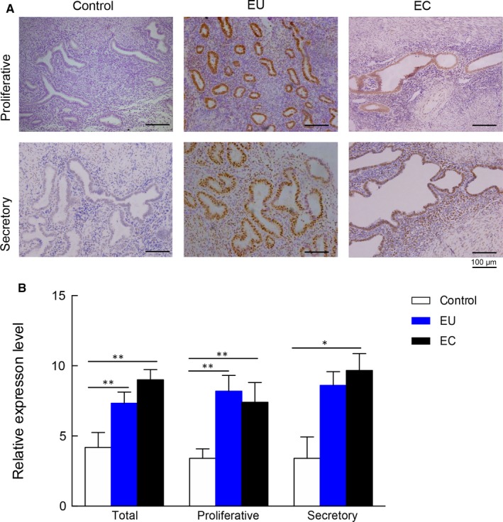
Representative immunohistochemical staining micrograph of C/EBPβ in different endometrial tissues. A, The representative immunohistochemical staining of eutopic, ectopic endometrium and matched controls in proliferative and secretory phase (original magnification 200×). B, The statistical analysis results of immunohistochemical staining from 10 patients with adenomyosis and 10 matched controls. EC, ectopic endometrium; EU, eutopic endometrium; **P < .01, *P < .05