
Figure 1.
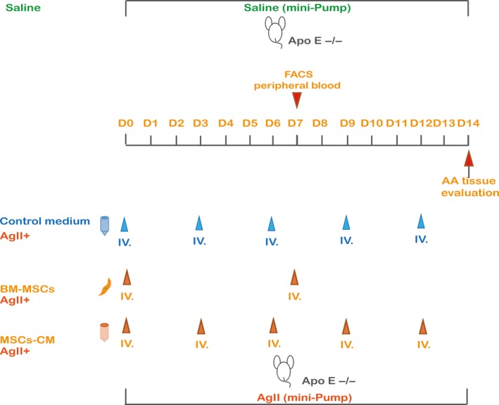
Schema of in vivo protocol. AA was induced by Ang II infusion for 2 wk in ApoE−/− mice; 1 × 10⁶ BM‐MSCs (in 0.2 mL saline) were transplanted via tail vein on day 0, and day 7. 0.2 mL MSCs‐CM or 0.2 mL DMEM alone was injected via the tail vein on day 0, day 3, day 6, day 9 and day 12. Peripheral blood was collected on day 7 for flow cytometry. Instead of AngII administration, saline was used as sham. All mice were sacrificed, and AA tissue was evaluated on day 14