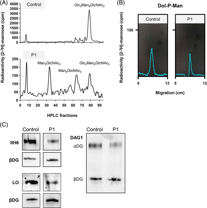
Figure 2.

CDG diagnostics and biochemical analyses of MPDU1‐CDG P1. A, HPLC analysis of LLO from fibroblasts of patient 1 (P1) and a control revealed the accumulation of the shortened dolichol‐linked oligosaccharides Man5GlcNAc2 and Man9GlcNAc2. B, Thin‐layer chromatography (TLC) analysis of hydrophobic LLO extracts further revealed that dolichol‐phosphate‐mannose (Dol‐P‐Man) is synthesized in patient 1 fibroblasts. C, Analysis of O‐mannosylated αDG in P1 and control fibroblasts. IIH6 and laminin (laminin overlay, LO) only bind to fully functional O‐mannosyl glycans of αDG. DAG1 binds to the core of the dystroglycan protein, showing expression of αDG and βDG proteins. CDG, congenital disorders of glycosylation; HPLC, High‐performance liquid chromatography; LLO, lipid linked oligosaccharide; MPDU1, mannose‐phosphate‐dolichol utilization defect 1
