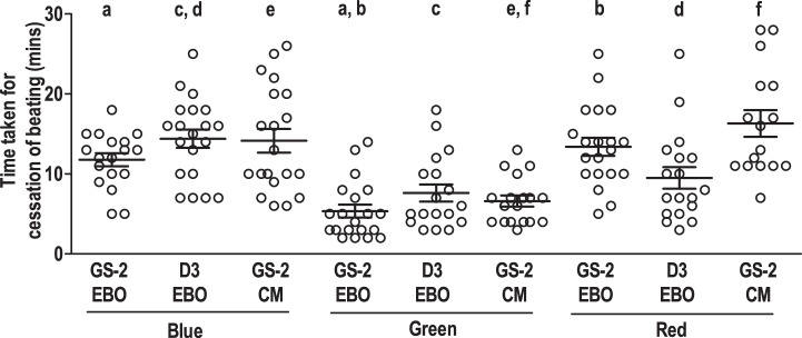
Fig. 4.
Time kinetics of cessation of cardiomyocyte contractility in beating clusters and enriched cardiomyocytes derived from embryonic stem (ES) cells, following exposure to monochromatic light. Time taken for cessation of cardiomyocyte contractility in beating clusters in intact EB outgrowths (EBO) derived from transgenic GS-2 ES cells, wild-type D3 ES cells, and enriched GS-2 ES cell-derived cardiomyocytes (CMs) following exposure to blue, green, and red light, respectively. Values represent means ± SE from 4 different experiments using 3–6 clusters in each experiment with a total of 14–20 clusters each group. Values with the same alphabet differ significantly: P < 0.0005 (f), 0.005 (a, b, c, and e), and 0.05 (d).

