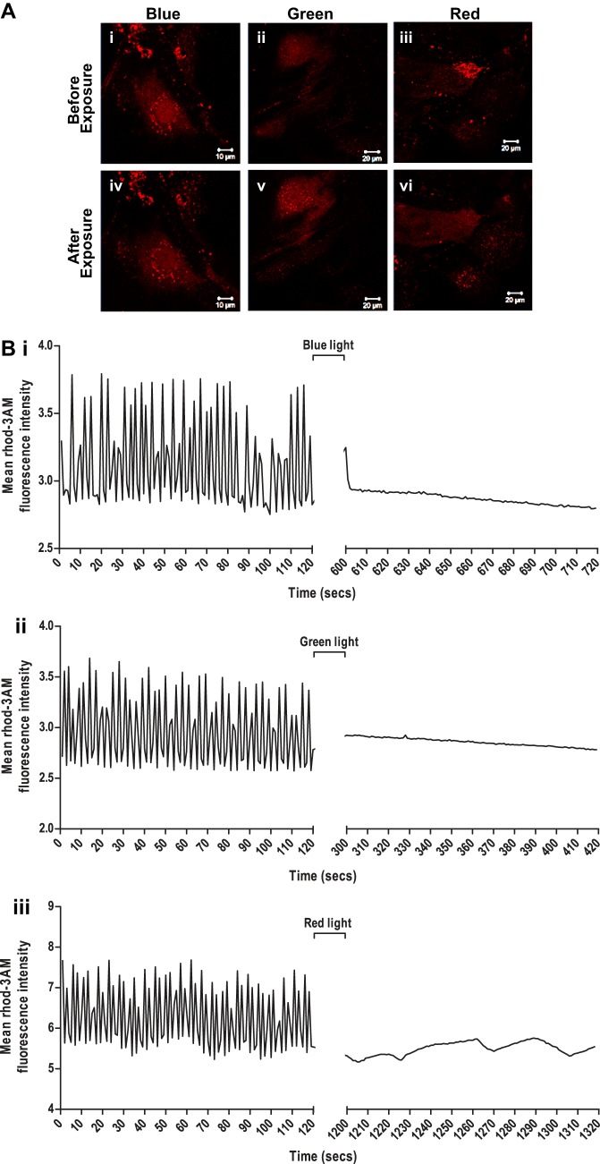
Fig. 7.
Assessment of intracellular calcium transients in enriched cardiomyocytes derived from GS-2 embryonic stem (ES) cells, following exposure to monochromatic light. A: intracellular calcium in enriched cardiomyocytes derived from GS-2 ES cells before (i-iii) and following (iv-vi) exposure to blue (i and iv), green (ii and v), and red (iii and vi) light visualized by rhod 3-AM staining. Scale bar: 10 µm (i and iv) and 20 µm (ii, iii, v, and vi). B: quantification of intracellular calcium transient-enriched cardiomyocytes derived from GS-2 ES cells before and after exposure to blue (i), green (ii), and red (iii) light measured by changes in relative rhod 3-AM fluorescence intensity in the cells.

