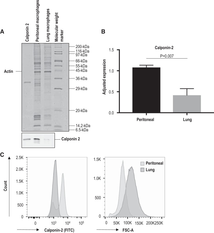
Fig. 1.
Significantly lower level of calponin 2 in lung macrophages than in peritoneal macrophages. A: the levels of calponin 2 in lung and peritoneal macrophages isolated from wild-type mice (n = 4 mice) were determined by immunoblotting. The protein input was normalized by the level of actin. Purified mouse calponin 2 protein was used as a positive control. B: the results were quantified by densitometry analysis using ImageJ software. The level of calponin 2 expression was evaluated relative to the levels of histones that reflect the number of cells. Bars depict means ± SE. C: the expression of calponin 2 in CD45+F4/80+ macrophages was confirmed by flow cytometry using rabbit anti-calponin 2 antibody RAH2 and FITC-conjugated anti-rabbit IgG. The rightward shift in forward scatter area (FSC-A) demonstrates the larger size of freshly isolated lung macrophages when compared with that of peritoneal macrophages.

