Fig. 7.
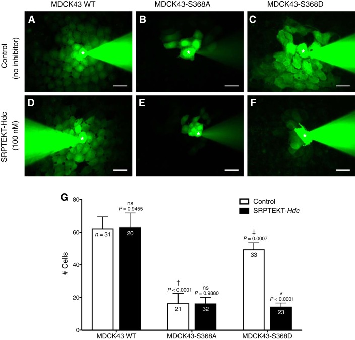
Dye coupling is blocked by SRPTEKT-Hdc in a S368 phosphorylation-dependent manner. A–F: representative images of NBD-MTMA dye coupling in MDCK43 WT cells (A and D); MDCK43-S368A cells (B and E); and MDCK43-S368D cells (C and F). Cells in A, B, and C were untreated, and those in D, E, and F were treated for 61–105 min with 100 nM SRPTEKT-Hdc before dye injection. Images were taken following 5 min of continuous injection; pipettes are evident in each image. Asterisks in fluorescent images denote microinjected cell. Note that coinjected rhodamine dextran did not diffuse to neighboring cells (not shown). Scale bar = 30 µm. G: number of cells receiving dye for each treatment condition. White bar, absence of SRPTEKT-Hdc; black bars, presence of SRPTEKT-Hdc. Values are means ± SE (sample sizes are indicated in each bar). P values comparing controls across cell types are from ANOVA and are indicated above the bars: †significant decrease in dye coupling compared with dye coupling in untreated MDCK43 WT cells; ‡significant increase in dye coupling in MDCK43-S368D cells compared with dye coupling in MDCK43-S368A cells. P values comparing untreated to peptide-treated are from t-test: *significant reduction in dye coupling compared with dye coupling in the absence of agent. ns indicates no significant difference in wave propagation.

