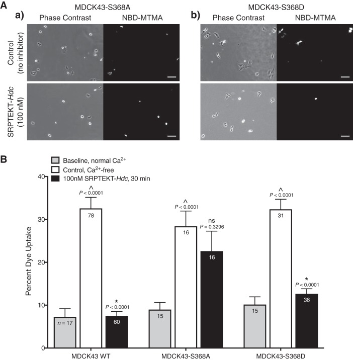
Fig. 8.
Dye uptake is blocked by SRPTEKT-Hdc in a phosphorylation-dependent manner. NBD-MTMA dye uptake under Ca2+-free conditions is shown. A: representative phase-contrast and NBD-MTMA fluorescence images of MDCK43-S368A cells (a) or MDCK43-S368D cells (b) under untreated, control conditions or following 15-min pretreatment in 100 nM SRPTEKT-Hdc followed by 15-min dye uptake in the presence of 100 nM SRPTEKT-Hdc. Rhodamine images are not shown as no cells contained this dye. Scale bar = 100 µm. B: percentage of NBD-MTMA-positive MDCK43 WT [as previously published (9)], MDCK43-S368A and MDCK43-S368D cells under baseline (normal Ca2+) conditions, control (untreated, Ca2+-free) conditions, and following exposure to 100 nM SRPTEKT-Hdc. The mean (± SE) percentage of NBD-MTMA-positive cells is shown (sample sizes are indicated in each bar). P values (from t-test) are indicated above the bars: ^significant increase in dye uptake compared with baseline; *significant reduction in percent dye uptake compared with the absence of agent.

