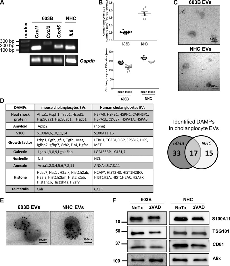
Fig. 1.
Human and mouse cholangiocytes release S100A11-containing extracellular vesicles (EVs). A: RNA from 603B mouse cholangiocyte cell line and normal human cholangiocyte (NHC) cell line was extracted and subjected to conventional/qualitative RT-PCR for Cxcl1, Cxcl2, and Cxcl5 gene expression (mouse) or IL8 gene expression (human). Gapdh was used as housekeeping gene. Gapdh in NHC was run on a separate gel. B: EVs were isolated from 603B and NHC cultures by differential ultracentrifugation and analyzed by nanoparticle tracking analysis. Concentration (top) and size distribution (bottom) are shown (603B n = 12; NHC n = 6, from independent EV preparations). C: transmission electron microscopy (TEM) images of 603B and NHC EVs. D: list of damage-associated molecular patterns (DAMPs) identified by liquid chromatography/tandem mass spectrometry on EVs isolated from 603B and NHC culture media. E: representative TEM images of 603B and NHC EVs following immunogold labeling with an anti-S100A11 antibody. F: immunoblot analysis for EV markers and S100A11 in 603B and NHC EVs after incubation with or without (NoTx) the pan-caspase inhibitor zVAD-fmk (10 μM) for 24 h.

