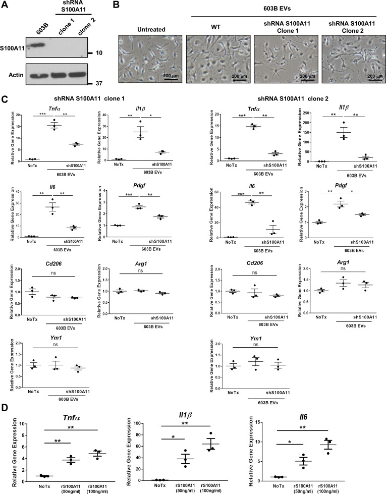
Fig. 2.
Cholangiocyte-derived extracellular vesicles (EVs) induce S100A11-mediated proinflammatory macrophage polarization. A: immunoblot analysis for S100A11 of cell lysates from 603B and S100A11 shRNA 603B (clone 1 and 2) cells. Actin was used as loading control. B: representative phase contrast microphotographs of wild-type mouse bone marrow-derived macrophages (BMDM) incubated without (untreated) or with EVs purified from the culture media of 603B and shRNA S100A11 603B cells (clone 1 and 2) at a concentration of 109/mL for 6 h. WT, wild type. C: expression of proinflammatory (Tnfα, Il1β, Il6), profibrogenic (Pdgf), and anti-inflammatory genes (Cd206, Arg1, Ym1) analyzed by quantitative (q)RT-PCR in BMDM treated as in B (n = 3, from independent BMDM and EV isolations). Target gene expression normalized to 18s. D: proinflammatory gene expression in wild-type BMDM treated with recombinant mouse S100A11 at the indicated concentrations for 6 h by qRT-PCR (n = 3, from independent BMDM isolations). Target gene expression normalized to 18s. Data represent mean of fold increase over control (untreated BMDM) ± SE. Two-tailed Student’s t-test: *P < 0.05, **P < 0.005, ***P < 0.001.

