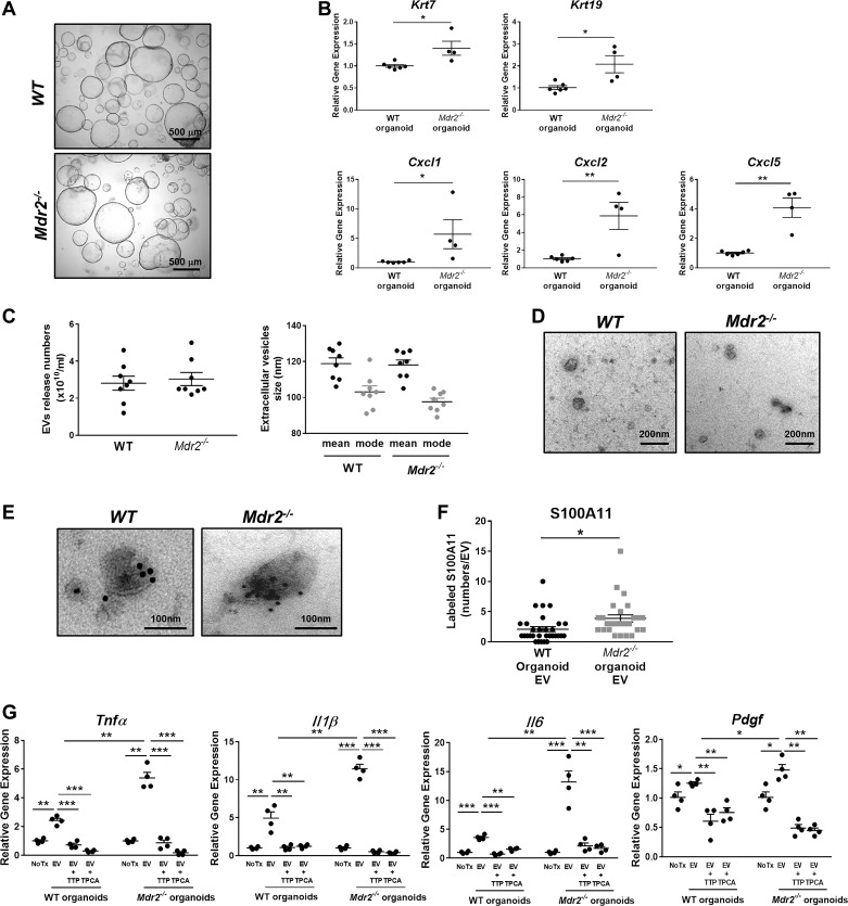
Fig. 5.
Extracellular vesicles (EVs) from Mdr2−/− mouse cholangiocyte-derived organoids promote proinflammatory polarization in bone marrow-derived macrophages (BMDM). A: representative phase-contrast photomicrographs of wild-type (WT) and Mdr2−/− mouse cholangiocyte organoids (×4). B: Krt7, Krt19, Cxcl1, Cxcl2, and Cxcl5 gene expression by quantitative RT-PCR in WT and Mdr2−/− mouse cholangiocyte-derived organoids at passage 1 (WT n = 6; Mdr2−/− n = 4, from independent isolations). Target gene expression normalized to Hprt and expressed as fold increase over WT cholangiocyte-derived organoids. Data represent mean of fold increase over control (WT) ± SE. Two-tailed Student’s t-test: *P < 0.05, **P < 0.005. C: concentration (left) and size distribution (right) of EVs isolated from WT and Mdr2−/− mouse cholangiocyte-derived organoids analyzed by nanoparticle tracking analysis (n = 8, from independent isolations). D: transmission electron microscopy (TEM) images of EVs from WT and Mdr2−/− mouse cholangiocyte-derived organoids. E: representative TEM images of EVs from WT and Mdr2−/− mouse cholangiocyte-derived organoids following immunogold labeling with an anti-S100A11 antibody. F: quantification of number of S100A11-positive particles on EVs from WT and Mdr2−/− mouse cholangiocyte-derived organoids (10 EV/genotype from 3 independent isolations). *P < 0.05. G: gene expression analysis by quantitative RT-PCR in WT BMDM treated with EVs from WT and Mdr2−/− mouse cholangiocyte-derived organoids in the presence or absence of TPCA-1 or TTP448 for 6 h. Target gene expression was normalized to Hprt. Data represent mean of fold increase over control (untreated BMDM) ± SE. Two-tailed Student’s t-test: *P < 0.05, **P < 0.005, ***P < 0.001 (n = 4, from independent EV isolations).

