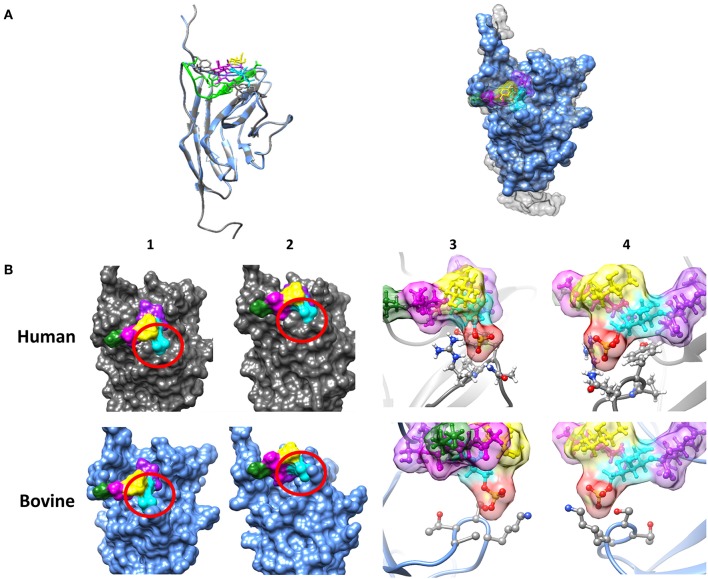
Figure 2.
Investigation of the sialic acid binding properties of human and bovine Siglec-8. (A) Overlay of the tertiary structure of hSiglec-8 (gray) and bovine Siglec-8 (blue). The components of the glycan are differently labeled: sialic acid (purple), fucose (yellow), galactose-6-P (cyan), N-acetylgalactosamine (magenta), 3-aminopropan-1-ol (green). (B) Differences in the tertiary structure of hSiglec-8 (gray) and bovine Siglec-8 (blue). Different views of the glycan-binding domain are shown (1–4). (3,4) 6-P is labeled in red. All figures were created by Chimera 1.13.1. Red circle demonstrates potential differences in glycan-binding properties. The structure of hSiglec-8 is based on Pröpster et al. (41) and the structure of bovine Siglec-8 was designed using Phyre2.

