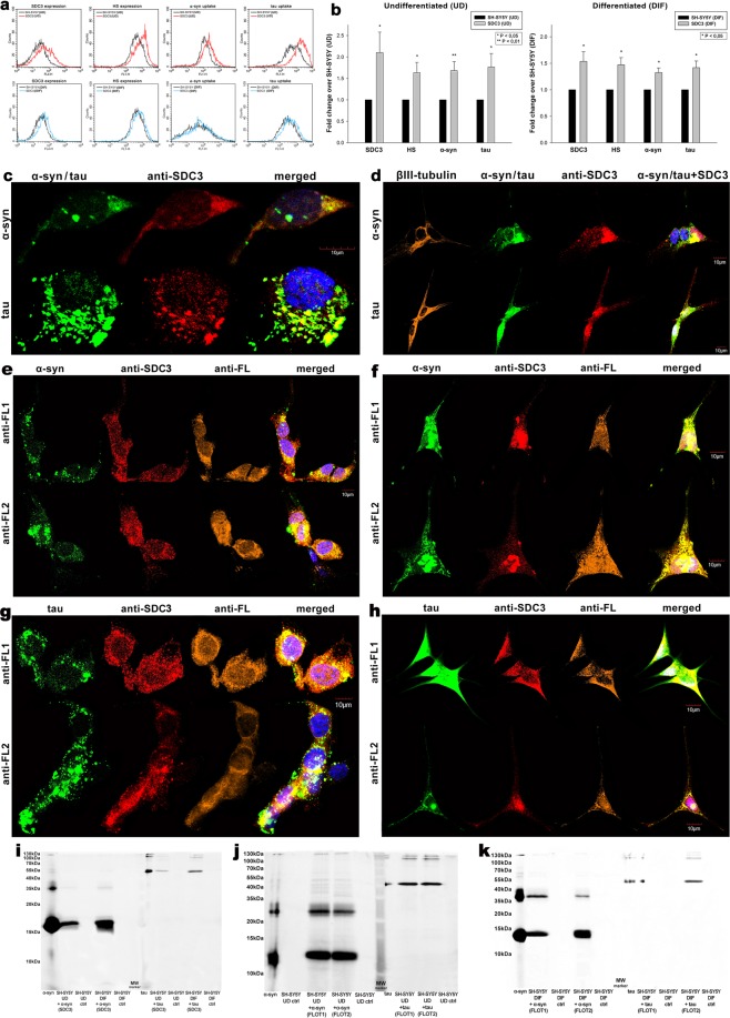
Figure 4.
Effect of SDC3 overexpression on α−syn and tau fibril uptake in SH-SY5Y cells. SDC3 transfectants, created in either differentiated (DIF) or undifferentiated (UD) SH-SY5Y cells, were selected by measuring SDC3 expression with flow cytometry using APC-labeled anti-SDC3 antibody. HS expression of SDC3 transfectants, along with WT SH-SY5Y, was also measured with flow cytometry using anti-HS antibody. SDC3 transfectants and WT SH-SY5Y cells were treated with FITC-labeled α−syn and tau fibrils (at a concentration of 5 µM monomer equivalent) at 37 °C. Cells incubated with fluorescent α−syn and tau fibrils for 3 h were then processed for uptake studies. (a) Flow cytometry histograms representing SDC3, HS expression levels and intracellular fluorescence of WT SH-SY5Y cells and SDC3 transfectants treated with fluorescent α−syn or tau fibrils. (b) Fold change in SDC3 and HS expression, along with α−syn and tau uptake following SDC3 overexpression in undifferentiated (UD) or differentiated (DIF) SH-SY5Y cells. The bars represent mean ± SEM of six independent experiments. Statistical significance vs fibril (α−syn or tau) treated WT SH-SY5Y cells as standards was assessed by analysis of variance (ANOVA). *p < 0.05 vs fibril (α−syn or tau) treated WT SH-SY5Y cells as standards. (c,d) Colocalization of the fibrils with SDC3 in undifferentiated (c) or differentiated (d) SH-SY5Y cells. CLSM images of WT SH-SY5Y cells treated with either of the FITC-labeled fibrils (α−syn or tau), along with APC-labeled SDC3 antibody. In case of differentiated SH-SY5Y cells (DIF), neuronal differentiation was justified by staining the cells with neuron-specific human βIII-tubulin antibody and Alexa Fluor 546-labeled secondary antibody. Scale bar = 10 μm. (e–h) Colocalization of the fibrils with flotillins undifferentiated (e,g) or differentiated (f,h) SH-SY5Y cells. CLSM images of WT SH-SY5Y cells treated with either of the fluorescent fibrils (α−syn or tau), along with APC-labeled SDC3 antibody and either of the Alexa Fluor 546-labeled FLOT1 and FLOT2 antibodies. Scale bar = 10 μm. (i–k) SDS-PAGEs showing fluorescent α−syn or tau immunoprecipitated with either SDC3 or one of the flotillin antibodies from extracts of undifferentiated (UD) or differentiated (DIF) SH-SY5Y cells.

