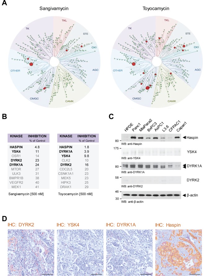
Figure 2.
Kinases targeted by Sangivamycin and Toyocamycin. (A,B) KINOMEscan screening was used to identify the kinase specificity of Sangivamycin and Toyocamycin at a concentration of 500 nM. The top ten hits for each compound is listed in the tables in. (B) The four overlapping kinases are highlighted in bold. (C) Cell lysates of HPDE cells or indicated PDA cell lines were analyzed by Western blot for expression of the main kinases targeted by Sangivamycin and Toyocamycin (anti-Haspin, anti-YSK4, anti-DYRK1A, anti-DYRK2). Staining for β-actin served as control for equal loading. (D) Immunohistochemistry (IHC) analysis of human PDA samples using anti-DYRK2, anti-YSK4, anti-DYRK1A, and anti-Haspin antibodies. The bar represents 100 µm.

