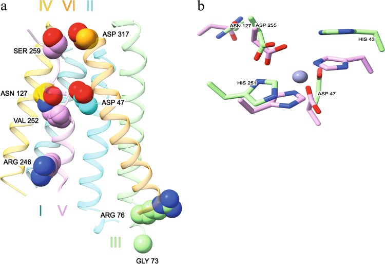
Fig. 3. The structure model of ZnT1 and the implications of inactivating missense mutations.
a Structural prediction of the transmembrane regions of ZnT1, using RaptorX alignment, and then the MEMOIR modeling server, with the bacterial Shewanella oneidensis zinc transporter 3j1z as a template. The residues selected for functional validation are represented as spheres. The roman numerals signify the number of the TM helix. b Structural superposition of zinc-binding site A in ZnT1 (green residues) and ZnT2 (violet residues), with a zinc ion (purple) from the YiiP structure

