
Fig. 2.
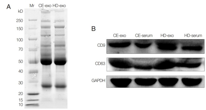
Protein analysis of CE-exo and HD-exo. (A) SDS-PAGE and Coomassie Blue stained. (B) Expression of CD9 and CD63 analyzed with Western blot using antibodies against CD9 and CD63. GAPDH was used as a normalizing control.
Official websites use .gov
A
.gov website belongs to an official
government organization in the United States.
Secure .gov websites use HTTPS
A lock (
) or https:// means you've safely
connected to the .gov website. Share sensitive
information only on official, secure websites.
Protein analysis of CE-exo and HD-exo. (A) SDS-PAGE and Coomassie Blue stained. (B) Expression of CD9 and CD63 analyzed with Western blot using antibodies against CD9 and CD63. GAPDH was used as a normalizing control.