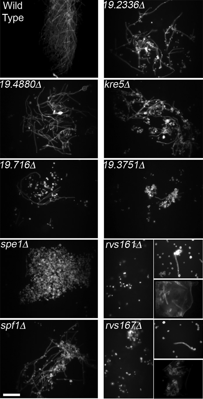
FIG 4.

Visualization of C. albicans cell morphology in tongue homogenates. In order to gain a broader representation of C. albicans morphology, infected tongues were homogenized and treated with 1 M KOH to dissolve mouse tissue, and the C. albicans cell walls were then stained with the fluorescent dye calcofluor white and visualized by fluorescence microscopy. The mutants are ordered according to the extent of weight loss that they caused in mice due to OPC (Fig. 2A). The strains are described in Table S2 in the supplemental material. Similar results were obtained in at least two independent infection experiments. Bar, 50 μm.
