Figure 1.
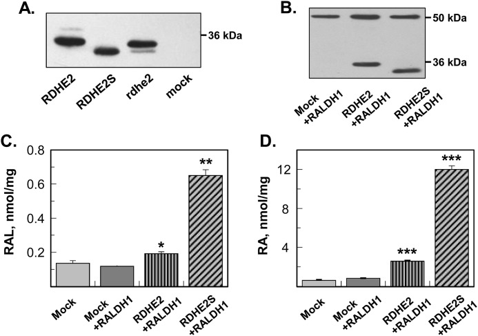
Expression and characterization of frog and murine RDHE2 and RDHE2S. A, Western blot analysis of microsomal fractions (5 μg) isolated from control Sf9 cells (mock) or cells expressing murine RDHE2, murine RDHE2S, or Xenopus rdhe2. His tag antibodies were used at a 1:1,000 dilution. B, Western blot analysis of HEK293 cell lysates (20 μg) containing murine RDHE2 or RDHE2S co-expressed with human RALDH1. RDHE2 and RDHE2S were detected using FLAG tag antibodies at a 1:3,000 dilution. RALDH1 was detected using HA antibodies at a 1:50 dilution. C, HPLC analysis of all-trans-retinaldehyde (RAL) production from all-trans-retinol (10 μm). Cells were incubated for 11 h. D, HPLC analysis of RA production from all-trans-retinol (10 μm). Samples are as indicated. Mock, cells transfected with empty vector. *, p < 0.01; **, p < 0.001; ***, p < 0.0001; mean ± S.D. (error bars), n = 3.

