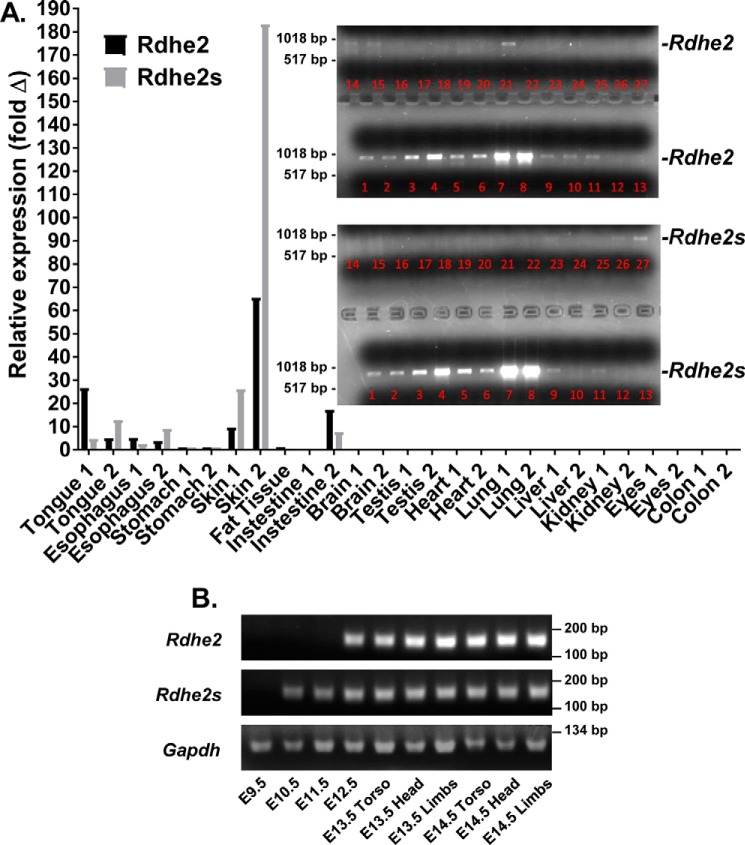
Figure 2.
Expression profile of Rdhe2 and Rdhe2s transcripts in mouse tissues. A, qPCR analysis of Rdhe2 and Rdhe2s expression in adult tissues of two animals. Inset, PCR-amplified full-length Rdhe2 and Rdhe2s transcripts. Lanes 1 and 2, tongue; lanes 3 and 4, esophagus; lanes 5 and 6, stomach; lanes 7 and 8, skin; lane 9, adipose tissue; lanes 10 and 11, intestine; lanes 12 and 13, brain; lanes 14 and 15, testis; lanes 16 and 17, heart; lanes 18 and 19, lung; lanes 20 and 21, liver; lanes 22 and 23, kidney; lanes 24 and 25, eyes; lanes 26 and 27, colon. B, semiquantitative PCR analysis of temporal expression patterns of Rdhe2 and Rdhe2s transcript fragments in embryonic mouse tissues. C57BL/J6 embryos were collected at E9.5–E14.5 stages of development as indicated. Five μg of mRNA was used for reverse transcription with random hexamer primers, and one-twentieth of the resulting cDNA was used per reaction. Gapdh was used as a loading control.

