
Figure 1.
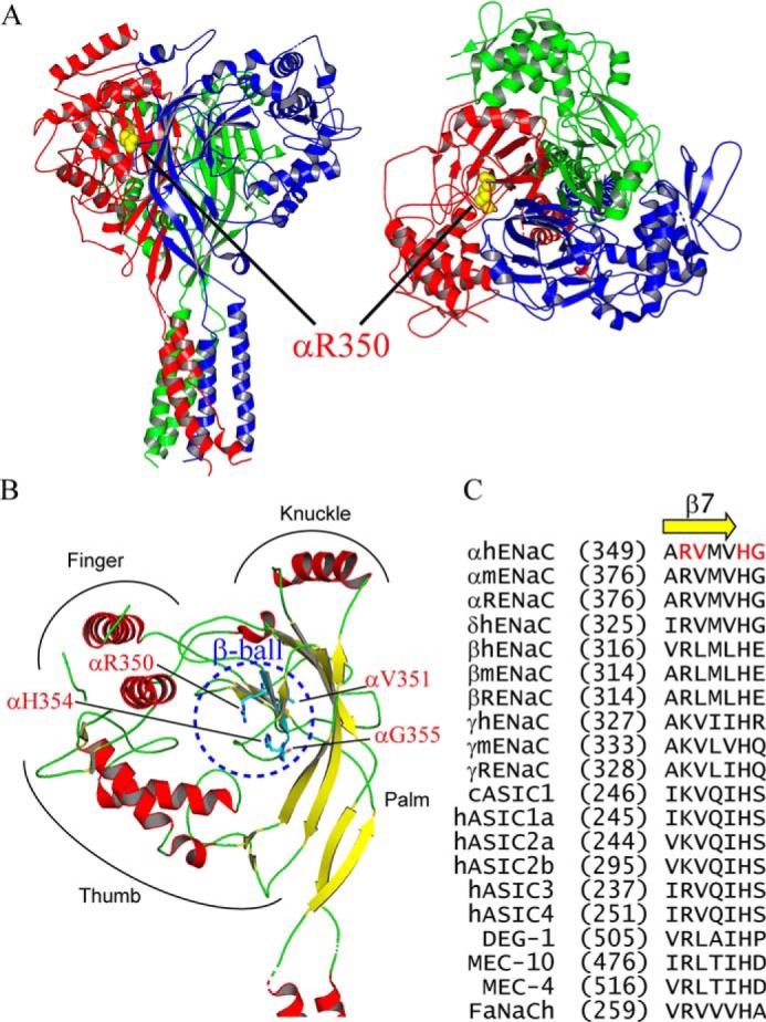
Location of variants in the human ENaC structure. A, location of αArg-350 in a trimeric human ENaC model (PDB 6BQN (21)). The α, β, and γ-subunits are shown as red, blue, and green ribbons, respectively, using PyMOL 2.0 (60). Side chain of αArg-350 is shown as yellow spheres. Left, side view, and right, top view. Both views show that αArg-350 is near the palm domain of βENaC. B, locations of αArg-350, αVal-351, αHis-354, and αGly-355 in the extracellular β-ball domain of αENaC. Helical domains are displayed in red and β-strands in yellow. All four residues are shown as sticks with carbons in cyan, oxygen in red, and nitrogen in blue. C, sequence alignments of ENaC/degenerin members. Alignments were performed using Vector NTI 11 (Thermo Fisher Scientific). Only sequences of the β7-strand and its following residues are shown. Amino acid numbers of the first residue in all sequences are shown in parentheses. Four residues where variants of this study reside are shown in red letters.