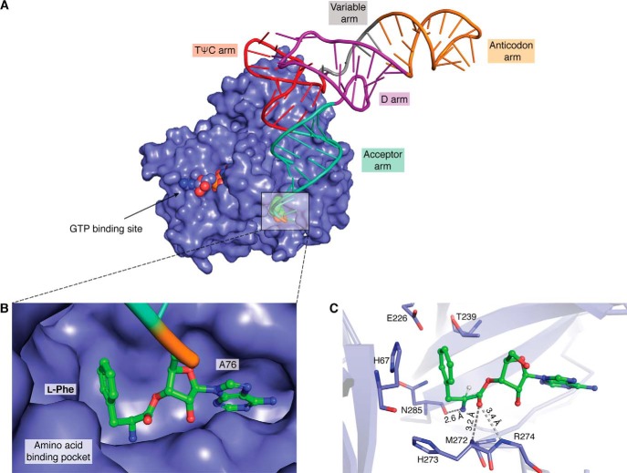
Figure 4.
Elongation factor in complex with l-Phe-tRNAPhe. A, crystal structure of a ternary complex, EF-Tu (surface representation in blue), GTP (shown as spheres in the GTP-binding site), and l-Phe-tRNAPhe (tRNA shown in a wire and stick representation with amino acids as spheres) (PDB entry 1TTT). B, zoomed in view of an amino acid (of l-Phe-tRNAPhe) bound in the amino acid–binding pocket of EF-Tu. C, stick representation of the amino acid–binding pocket showing key interactions with the ligand (l-Phe).

