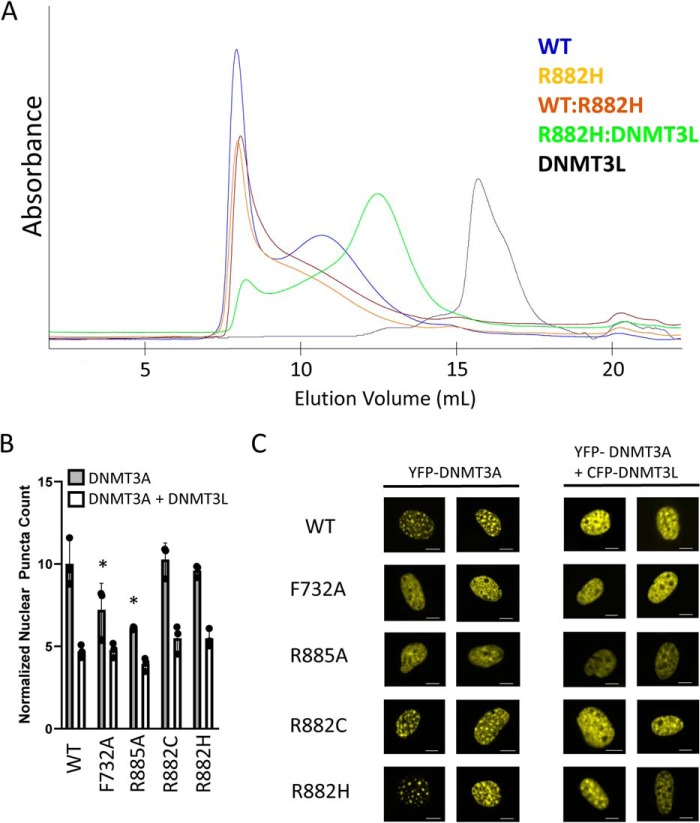
Figure 3.
Examining the oligomeric state of R882H DNMT3A. A, size-exclusion chromatograms of DNMT3A and DNMT3L proteins. Mixed complexes were pre-incubated for 2 h at room temperature prior to injection onto a Superose 6 column. Peak components were confirmed by SDS-PAGE analysis. 500 μg of each protein was injected. B, quantification of nuclear puncta in NIH-3T3 cells after transfection with YFP–DNMT3A with or without co-transfection of CFP–DNMT3L. Error bars represent S.D. F732A and R885A have a statistically significant lower number of nuclear puncta compared with WT, p < 0.05 (*). There is no difference in the number of mutant DNMT3A nuclear puncta compared with WT upon DNMT3L co-expression. C, representative images are from B. Scale bars, 5 μm.

