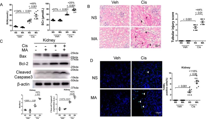
Figure 2.
MA deteriorated renal function and enhanced apoptosis in cisplatin-treated kidneys. Blood and kidney samples were collected 3 days after cisplatin administration. A, BUN and serum creatinine (SCr) levels. B, hematoxylin–eosin staining was performed, and representative pathological images (original magnification, ×400; bar, 100 μm) of kidneys are shown. #, renal tubular cast; arrowhead, dilated renal tubule. Scale bar, 100 μm. Tubular injury was semiquantitatively scored. C, the expression of Bcl-2, Bax, and Cleaved Caspase-3 in kidneys was assessed by Western blotting and then quantitatively analyzed. D, TUNEL staining was performed to measure apoptosis in the kidney tissue sections (original magnification, ×400; bar, 100 μm). The data represent means ± S.D., and the results are representative of three independent experiments. Veh, vehicle; Cis, cisplatin; NS, normal saline.

