Figure 5.
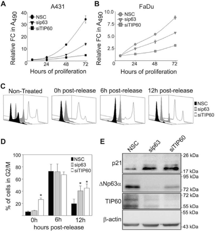
ΔNp63α and TIP60 silencing inhibits SCC proliferation. A and B, A431 (A) and FaDu (B) cells were transfected with NSC, sip63, or siTIP60 as indicated, and cell proliferation was measured by MTS cell titer. The y axis represents the -fold change of each condition relative to t = 6 h. Error bars represent standard deviation from the mean. C, cell cycle profiles of A431 cells transfected with NSC, sip63, or siTIP60 at the indicated time points after release following a double thymidine block and analyzed using flow cytometry. D, quantitation of the total percentage of cells progressing through G2/M 0, 6, and 12 h post-thymidine release under each condition shown in C. Error bars represent 1 S.E. of the mean from three independent experiments. *, p ≤ 0.05). E, immunoblot analysis of p21, ΔNp63α, and TIP60 of A431 cells (C) was performed with the indicated antibodies 12 h after release from double thymidine block.

