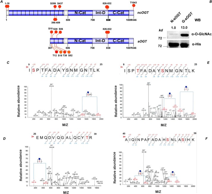
Figure 1.
Site mapping of O-GlcNAcylation on sOGT. A, known O-GlcNAcylation sites on sOGT/ncOGT. B, O-GlcNAc sOGT enriched through WGA affinity chromatography was examined via Western blotting. The blots were quantified with ImageJ, and O-GlcNAcylation of sOGT was normalized to the input (α-His). C–F, O-GlcNAc sOGT was expressed in E. coli cells, pooled with WGA beads, and digested in-gel with trypsin. The resulting peptides were analyzed on an LTQ-Orbitrap Elite mass spectrometer employing ETD fragmentation. ETD spectra revealed 1–2 O-GlcNAc modifications on each of the following peptides: ISPTFADAYSNMGNTLK (C), ISPTFADAYSNMGNTLK (D), EMQDVQGALQCYTR (E), and AIQINPAFADAHSNLASIHK (F) (all of the modification sites shown in underlined boldface letters).

