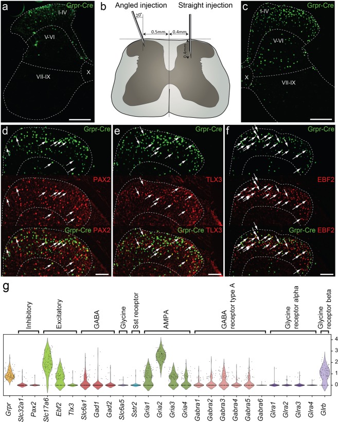
Figure 1.
The Grpr-Cre population of spinal interneurons is predominantly excitatory. Analysis of adult Grpr-Cre neurons displayed through a viral reporter system. (a) Coronal section of lumbar spinal cord annotated using Rexed laminae following an injection of AAV2-EF1a-DIO-EYFP virus using the angled injection scheme. Grpr-Cre neurons in green. (b) Illustrating the difference between the angled and the straight injection schemes. (c) Coronal section of lumbar spinal cord following an injection of the reporter virus AAVDJ-EF1a-DIO-HTB using the straight injection scheme. Grpr-Cre neurons in green. (d–f) Immunohistochemistry against PAX2 (d), TLX3 (e) and EBF2 (f) with Grpr-Cre neurons in green and the antigen in red. Dashed lines outline lamina I-IV. Arrows indicate Grpr-Cre neurons overlapping with the antigen. (g) Grpr neurons express both excitatory and inhibitory markers and genes important for glutamatergic, GABAergic and glycinergic signaling input. Violin plot of normalized expression of marker genes and AMPA, GABA receptor type A and glycine receptor subunits in Grpr-expressing neurons. For gene expression of all presented genes in all Häring cell types see Figure S3A. For gene expression in all Grpr neurons grouped by Häring cell types, see Figure S3B. Scale bars in (a) and (c) correspond to 200 µm while scale bars in (d–f) correspond to 100 µm.

