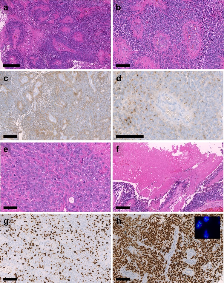
Fig. 2.
Histological features of SP-EPN-MYCN Highly cellular neuroectodermal tumor with a perivascular pseudorosettes, b microvascular proliferation, c perivascular enhanced GFAP expression, d dot-like EMA positivity, e brisk mitotic activity, f tumor necrosis, g high Ki-67 labelling, and h extensive nuclear MYCN expression. Inset in h): Results of two- color fluorescence in situ hybridization demonstrating multiple green signals for the MYCN-locus-probe and 2 red signals for the centromeric control probe. Scale bar = 300 µm in a, 100 µm in b, d, g, h, 200 µm in c, f and 50 µm in e

