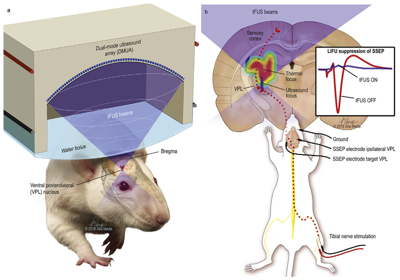
Fig. 1. Transcranial-focused low-intensity ultrasound neuromodulation.
Illustration of the delivery of transcranial focused ultrasound to the ventral posterolateral nucleus of the thalamus with the mechanical focus highlighted in blue and the larger and more proximate heating profile as a color gradient with maximal thermal delivery in red. Electrical stimulation of the tibial nerve produces signals that travel through the nucleus gracilis, followed by the medial lemniscus to the contralateral ventral posterolateral nucleus of the thalamus, which projects to the somatosensory cortex. Evoked somatosensory-evoked potentials (SSEP) from the VPL with and without ultrasound are illustrated (1b inset).

