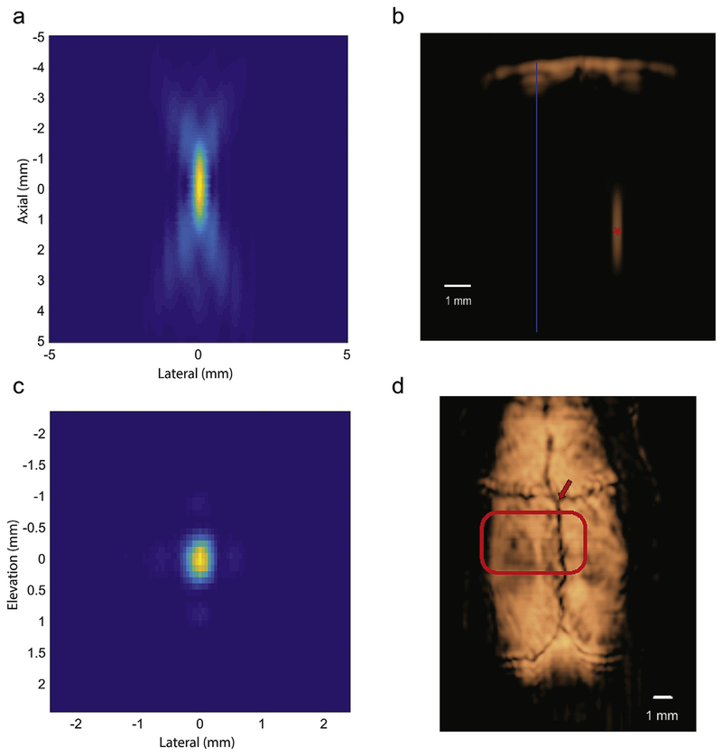
Fig. 2. Image-guided targeting and tFUS beam characterization.
a: Focus measured in degassed water demonstrating the axial extent of the focus (coronal section). b: B-mode image of the skull at 3 mm behind Bregma with the focal spot placement within the brain (6 mm deep) (red asterisk) with midline (blue). c: Focus measured in degassed water demonstrating the lateral-elevation profile (axial section). d: A C-scan image of a rodent skull surface from 3D DMUA imaging with the active ultrasound wavefront highlighted. The DMUA was positioned with respect to the intersection of the bregma and medial suture lines (arrow). The intersection of the bregma and medial suture lines (arrow) served as a marker for placing the DMUA for targeting the stereotactic coordinates. The highlighted region illustrates the cross section of the tFUS beam at the skull surface.

