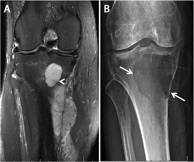
Figure 1.
Case 1, radiologic findings, preoperative and post neoadjuvant therapy. A) Preoperative coronal fat-saturated T2-weighted MRI image demonstrates heterogeneous hyperintensity in both intra- and extra-osseous components with a small perforating vessel at the posterior medial margin of the tibia (arrowhead) that may have allowed spread of the tumor between intraosseous and extraosseous components. The tumor vasculature in the soft tissue component is arborizing and hypointense. B) Radiograph of the tumor after neoadjuvant therapy shows a lytic lesion in the subarticular region of the proximal tibia, with sharp but non-sclerotic margins laterally, and a more poorly defined inferior margin. High-grade endosteal scalloping of the proximal tibia is present medially.

