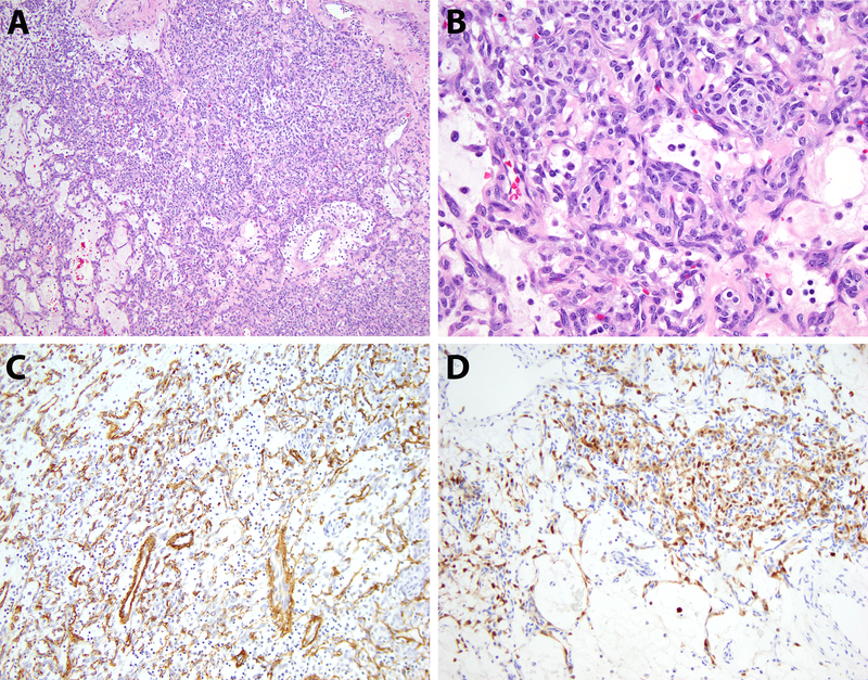
Figure 4.
Case 3, pathologic findings. A) Low-power view of the ovarian tumor shows neoplastic cells arranged predominantly in sheets with areas of alternating cellularity (100x, H&E). B) Cytologic features demonstrate bland-appearing ovoid tumor cells with scant cytoplasm and a vascular-rich stroma (400x, H&E). C,D) The tumor cells express SMA (focal) and S100 protein by immunohistochemistry (200x, immunohistochemical stains).

