Fig. 1.
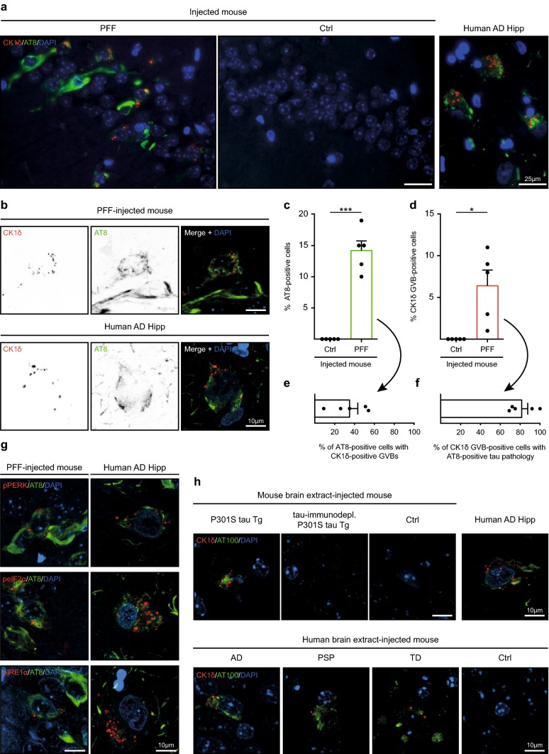
Seeding of tau pathology induces GVBs in vivo. a–g Immunofluorescence was performed on hippocampal sections of tau P301L Tg mice injected with K18 tau P301L seeds (PFF, N = 5) or control buffer (Ctrl, N = 5). a Representative epifluorescence images of immunostaining using AT8 (green) and the GVB marker CK1δ (red). Note that the AT8 epitope is absent from K18 PFFs. b Representative confocal images of neurons positive for immunolabeling with AT8 and CK1δ (shown separately in grayscale and in color in the merge: AT8 in green and CK1δ in red). c–f Quantification of a showing the percentage of pyramidal neurons positive for AT8 (c) or CK1δ (d) in the hippocampal CA1, CA2 and CA3 area of Ctrl or PFF-injected mice. *p < 0.05, ***p < 0.001, one-sample t test. e Quantification of the percentage of AT8-immunoreactive neurons that contains CK1δ-labeled GVBs and f of neurons with CK1δ-positive GVBs that is also positive for AT8 in the hippocampus of PFF-injected mice. Bars indicate the mean + SEM. Data points represent (mean values of) individual animals. See Supplementary Table 2 in Online Resource 1 for an overview of the number of cells analyzed. g Representative confocal images of neurons positive for immunostaining with AT8 (green) and the GVB markers pPERK, peIF2α and pIRE1α (red). h ALZ17 mice injected with extract from mouse brain [P301S tau Tg (N = 6), tau P301S Tg immunodepleted of tau (tau-immunodepl. P301S tau, N = 4), non-Tg C57BL/6 mice (Ctrl, N = 1)] or human post-mortem brain [AD (N = 6), PSP (N = 3), TD (N = 4) patients or control (Ctrl, N = 4)]. Representative confocal images of immunofluorescence with AT100 (green) and CK1δ (red) in the injected mice are shown. Human AD hippocampus (Human AD Hipp) is included for reference in all immunostainings. Cell nuclei are stained with DAPI (blue) in all images

