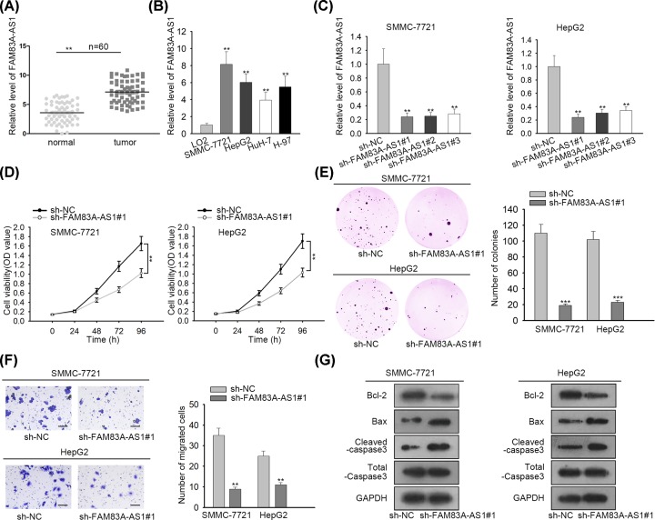
Figure 1. FAM83A-AS1 expression was elevated in HCC tissues and cells and deficiency of FAM83A-AS1 suppressed the progression of HCC.
(A) The expression of FAM83A-AS1 in HCC tissues and normal adjacent tissues were evaluated by qRT-PCR. (B) qRT-PCR detected the expression of FAM83A-AS1 in HCC cells and normal liver cells. (C) qRT-PCR was employed to test the knockdown efficiency of FAM83A-AS1 in HCC cells. (D) CCK-8 assay delineated cell proliferation ability in HCC cells. (E) Colony formation assay was carried out to examine the number of colonies in HCC cells. (F) Transwell assay was applied for estimating cell migration in HCC cells (scale bar = 200 μm). (G) Western blot assay was conducted to explore cell apoptosis in HCC cells. **P<0.01, ***P<0.001.

