Figure 2.

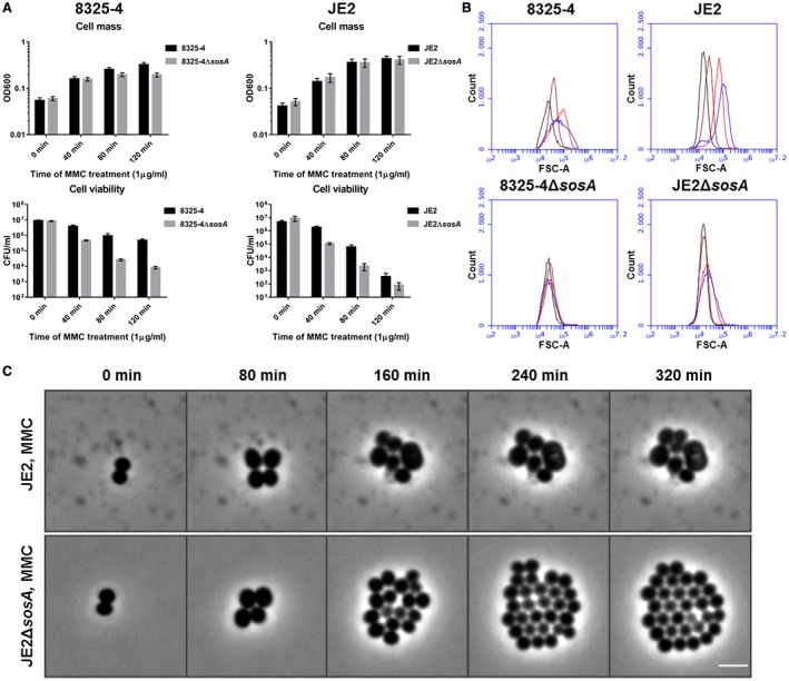
SosA supports survival of S. aureus subjected to lethal DNA damage and is involved in bacterial swelling. A. Culture optical density at 600 nm and cell viability of S. aureus strains 8325‐4 and JE2 in comparison with their respective ΔsosA mutants upon challenge with a lethal dose of mitomycin C (MMC, 1 µg ml−1) for 2 h. Error bars represent the standard deviation from three biological replicates. B. Cell size of 8325‐4 and JE2 WT and ΔsosA mutants exposed to mitomycin C estimated by flow cytometry (FSC‐A). Cells were grown exponentially prior to MMC addition at an OD600 of 0.05. Samples were taken after 0 (black), 40 (brown), 80 (red) and 120 (blue) min of incubation with MMC. C. Effect of MMC treatment (0.04 µg ml−1) on cell shape and cell number of JE2 WT and JE2ΔsosA as visualized by time‐lapse phase‐contrast microscopy. Scale bar represents 2 µm.
