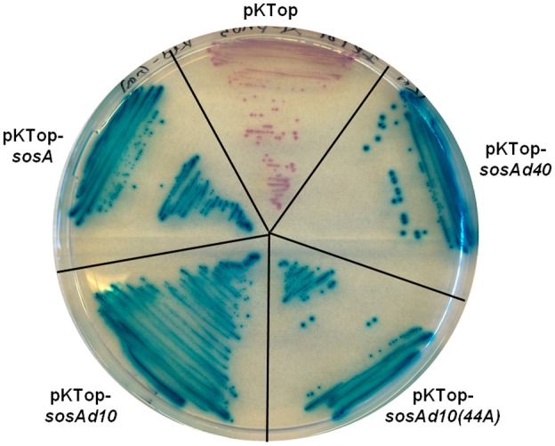
Figure 4.

Evaluation of membrane localization of SosA and truncated variants SosAd10, SosAd10(44A) and SosAd40 by in frame fusion at the C‐terminus to PhoA‐LacZ in pKTop (the truncated variants are explained in Figs 5 and 6). E. coli IM08B cells carrying the constructs were streaked on a dual‐indicator plate containing LB agar plus 50 µg−1 of kanamycin, 1 mM IPTG, 5‐bromo‐4‐chloro‐3‐indolyl phosphate disodium salt (80 µg ml−1) and 6‐Chloro‐3‐indolyl‐β‐D‐galactopyranoside (100 µg ml−1). Cytoplasmic localization of the PhoA‐LacZ chimera is indicated by red/rose color development whereas translocation across the membrane is indicated by blue color development.
