
Figure 4.
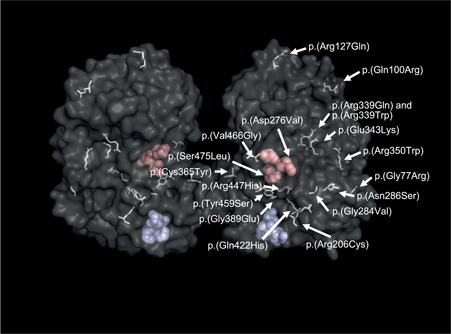
TPP1 proenzyme structure and missense variants reported ≥ three times. Three‐dimensional structure of TPP1 dimers (Pal et al., 2009). Active site (catalytic triad, Ser475‐Glu272‐Asp360) pocket residues are shown as red space‐filling models, calcium binding sites (Asp517‐Val518‐Gly539‐Asp543) in blue.