Figure 1.

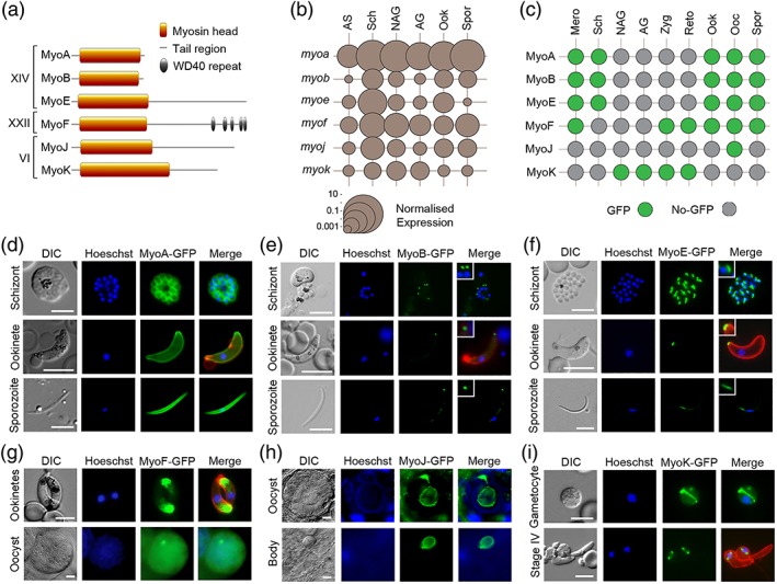
Expression and localisation of Plasmodium myosins throughout the life cycle. (a) There are six Plasmodium myosins. Two Class XIV myosins have a “head” and “neck” region but no tail. The remaining Class XIV and Class XXII and VI myosins have a “tail,” which in the case of MyoF contains WD40 repeat domains. (b) Plot of normalised transcript expression levels for each myosin gene throughout the Plasmodium life cycle. RNA was prepared from asexual blood stages (AS), blood stage schizonts (Sch), nonactivated gametocytes (NAG), activated gametocytes (AG), ookinetes (Ook), and sporozoites (Spr). Two genes, arginine‐tRNA synthetase and hsp70, were used as controls for normalisation. Each point is the mean of three biological replicates. See also Table S2. (c) Summary of GFP‐tagged myosin expression throughout the life cycle, in merozoites (Mero), schizonts (Sch), nonactivated gametocytes (NAG), activated gametocytes (AG), zygotes (Zyg), retort‐forms (Reto), ookinetes (Ook), oocysts (Ooc), and sporozoites (Spor). Expression of (d) MyoA‐GFP, (e) MyoB‐GFP, and (f) MyoE‐GFP in schizonts, ookinetes, and sporozoites using live cell imaging. MyoB and MyoE with higher magnification inset. Expression of (g) MyoF‐GFP in ookinetes and oocysts, (h) MyoJ‐GFP in an oocyst and residual oocyst body following sporozoite egression, and (i) MyoK‐GFP in a gametocyte and early (Stage IV) retort/ookinete using live cell imaging. Shown are differential interference contrast (DIC) image, Hoechst 33342 (blue), GFP (green), Merge (blue, green), and 13.1 (red), a cy3‐conjugated antibody recognising P28 (on activated female gametocytes, zygotes, and ookinetes only). Size marker = 5 μm
