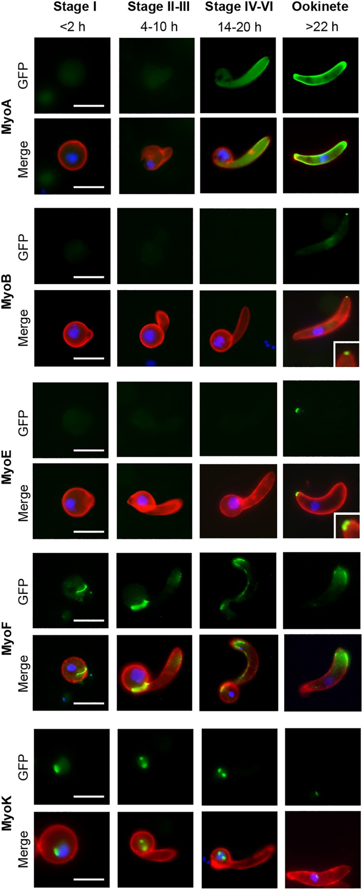
Figure 2.

Myosin expression during ookinete development. Localisation and expression of MyoA‐GFP, MyoB‐GFP, MyoE‐GFP, MyoF‐GFP, and MyoK‐GFP during ookinete development (Stage I, <2 hr; Stages II and III, 4–10 hr; Stages IV–VI, 14–20 hr; and mature ookinete, >22 hr) using live cell imaging. Top row: GFP (green), bottom row: Merge, Hoechst 33342 (blue); GFP (green); and 13.1 (red), a cy3‐conjugated antibody recognising P28 on the surface of activated female gametocytes, zygotes and ookinetes. MyoB and MyoE with higher magnification inset. Scale bar = 5 μm
