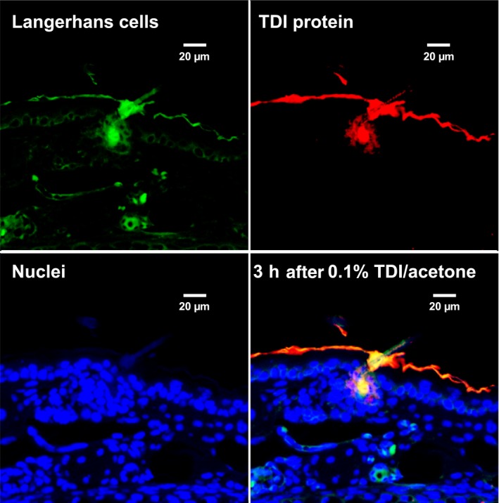
Figure 3.

Immunochemical co‐localization in mouse skin of 2,4‐TDI haptenated proteins (albumin, and cuticular and cytoskeletal keratins) along with antigen presenting cells (from: Nayak et al Tox Sci: 140(2) 327‐337, 2014). Dermal LMW chemical sensitization is often used with subsequent respiratory challenge to model LMW chemical asthma. TDI was observed to rapidly haptenate dermal proteins, especially in the outer root sheath of the hair follicle, and recruit antigen‐presenting cells (CD11b APCs, CD207 Langerhans cells and CD103+CD207+ Langerhans cells) with subsequent transport to local draining lymph nodes. Confocal microscopic images of Langerhans cells (top left), TDI haptenated tissue (top right), cell nuclei (bottom left) and overlay of Langerhans cells, nuclei, and TDI haptenated tissue (bottom right)
