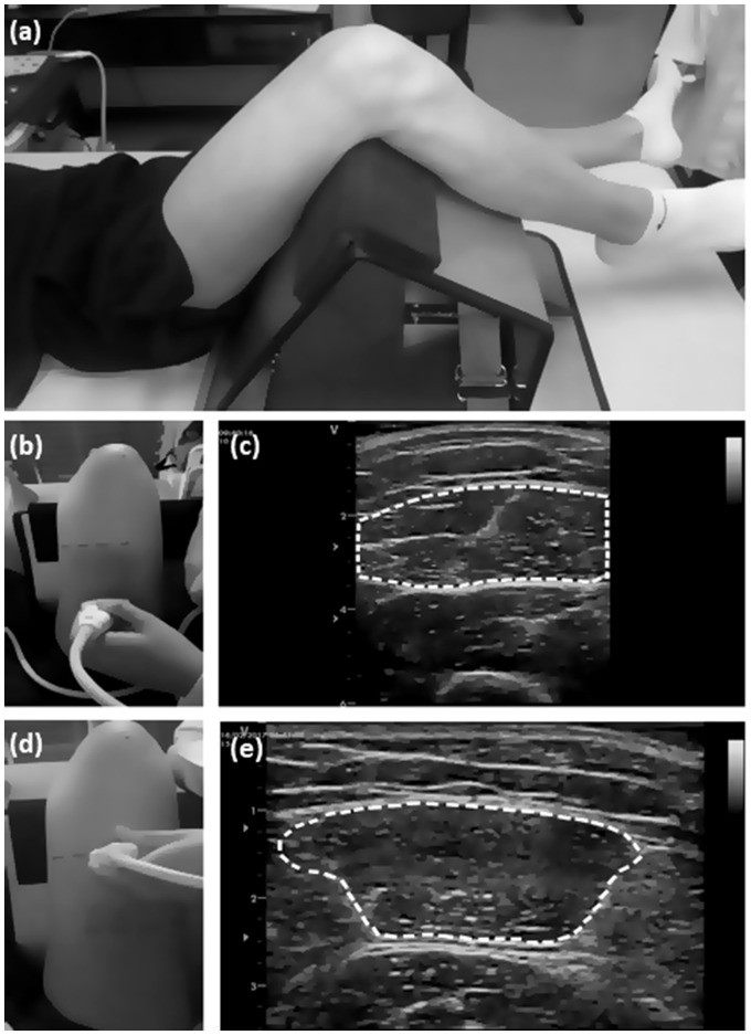
Figure 1.
Methodology used for ultrasound evaluation at the two rectus femoris (RF) muscle sites: (a) positioning of the subject for EI evaluation; (b) probe positioned at the RF50 site; (c) US image of the muscle on RF50 site; (d) probe positioned at the R70 site; (e) US image of the muscle on RF70 site. Dashed lines indicate how the EI area was determined at the ultrasound images.

