
Figure 2.
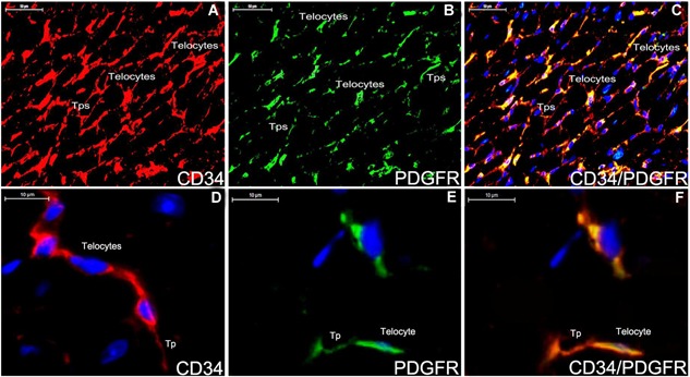
Double staining immunofluorescence for CD34/PDGFR‐α in the myocardium tissue of ApoE‐/‐ mice (A‐F). The distribution of telocyte is shown positively with CD34 (A,red), PDGFR‐α (B,green) and CD34/PDGFR‐α (C,yellow). Telocyte can construct a 3D network in the myocardium tissue (A‐C). The morphology of telocyte is completely shown (D‐F). CD34‐positive telocyte is oval‐liked with two telopodes (D,red). PDGFR‐α‐positive telocyte is spindle‐liked with one telopode (E,green). Telocyte expresses for CD34/PDGFR‐α (F,yellow). Nuclei were counterstained with DAPI(blue). The scale bar = 50μm (A‐C) and 10μm (D‐E).