Figure 3.

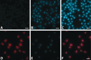
Images of Jurkat cells incubated for 15 mins with 4 at A) 50 nm, B) 0.5 μm, and C) 5 μm. Jurkat cells co‐stained with D) anti‐LCK‐AF647 and E) 4 (0.5 μm), with F) showing the overlay of the two channels. Scale bar: 25 μm.

Images of Jurkat cells incubated for 15 mins with 4 at A) 50 nm, B) 0.5 μm, and C) 5 μm. Jurkat cells co‐stained with D) anti‐LCK‐AF647 and E) 4 (0.5 μm), with F) showing the overlay of the two channels. Scale bar: 25 μm.