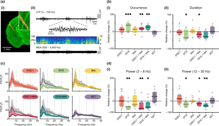
Figure 2.

Patterns of oscillatory activity in the i/vHP of neonatal mouse models of mental illness. (a) (i) Digital photomontage reconstructing the position of a 1‐shank DiI‐labelled recording probe (orange) in the HP of a Nissl‐stained 100 μm‐thick coronal section (green) from a P9 control mouse. Inset, the position of recording sites (white dots) over the Str. pyramidale displayed at higher magnification. (ii) Extracellular recording of discontinuous oscillatory activity in HP from a P9 control mouse displayed after band‐pass (3–100 Hz) filtering accompanied by the colour‐coded wavelet spectra at identical timescale (middle) and the corresponding MUA after band‐pass (500–5,000 Hz) filtering (bottom). Inset, discontinuous oscillatory event displayed at higher magnification. (b) (i) Scatter plot displaying the relative occurrence of oscillatory events in hippocampal CA1 area of all models when normalized to controls (Df16 vs. control: p = 0.001, DISC1 + MIA vs. control: p = 0.002, Df16 + MIA vs. control: p = 0.008). (ii) Same for the duration of oscillatory events (Df16 vs. control: p = 0.018, DISC1 + MIA vs. control: p = 0.018). (c) Averaged power spectra P(f) of discontinuous hippocampal oscillations normalized to the baseline power P 0(f) of time windows lacking oscillatory activity displayed for all mouse models (colours) together with their control (light grey). (d) (i) Scatter plot displaying the relative hippocampal power within 3–8 Hz for all models when normalized to controls (Df16 vs. control: p = 0.005, DISC1 + MIA vs. control: p = 0.004, Df16 + MIA vs. control: p = 0.043). (ii) Same as (i) in the beta (12–30 Hz) frequency band (Df16 vs. control: p = 0.018, DISC1 + MIA vs. control: p = 0.03, Df16 + MIA vs. control: p = 0.002). Thick lines represent the median, and shaded areas represent the 25° and 75° percentiles. Single data points are represented as circles, the coloured bars represent the median and the coloured boxes the 25th and 75th percentiles, *p < 0.05, **p < 0.01, ***p < 0.001
