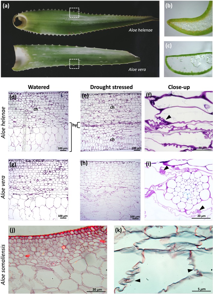
Figure 2.

Comparative morphology and anatomy of Aloe helenae (a, b, d, e, and f), Aloe vera (a, c, g, h, and i), and Aloe somaliensis (j and k). Transverse sections (marked by dashed squares) of hydrenchyma tissue stained with toluidine blue showing typical leaf tissue arrangement in Aloe species with hydrenchyma, (h) surrounded by an outer photosynthetic chlorenchyma, (c) epidermis and cuticle (d, e, g and h). Convoluted folding of hydrenchyma cell walls in drought‐stressed plants (f and i) indicated by arrows. Aloe somaliensis (j and k) from the Kew slide collection also showing folds in the hydrenchyma cell walls
