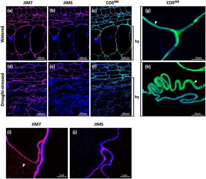
Figure 4.

In situ detection of homogalacturonan in sections of Aloe vera leaves using monoclonal antibodies JIM7 (binding highly methylated homogalacturonan), JIM5 (binding homogalacturonan with a low degree of esterification), and COS488 (binding highly deesterified homogalacturonan); images overlayed with Calcofluor White signal (blue) highlighting the cell walls. Micrographs showing (a–c) watered and (d–f) drought‐stressed specimens show remarkable changes in cell wall polysaccharide composition and folding. Detail is shown in (g–j) high magnification micrographs. Arrowheads indicate (g and h) middle lamella and (i) intracellular accumulation
