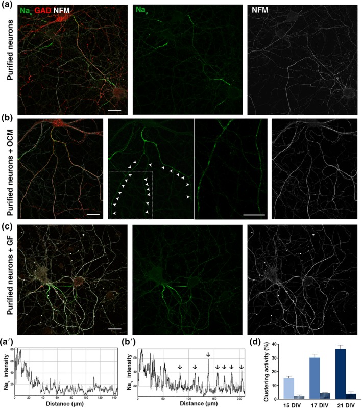
Figure 1.

OCM promotes Nav clustering on GABAergic hippocampal neurons in culture. (a, b, c) immunostainings of purified hippocampal neurons cultured in the absence of OCM (a), presence of OCM (b) or growth factors (GF); that is, IGF‐1, BDNF, and GDNF (c) fixed at 17 DIV. The axon initial segment (AIS) is detected in all conditions (a–c), but clusters of Nav (green) are only detected in the presence of OCM (b) on GABAergic axons (GAD67+; red). The periodic clusters of Nav channels are shown at a higher magnification of the framed part. Neurites are stained with an antibody targeting neurofilament M (NFM; white). Scale bars 25 μm. (a′, b′) Fluorescence intensity profiles correspond to axonal Nav immunolabeling from (a and b); individual peaks in b′ (arrows) represent Nav clusters. (d) Clustering activity represents the percentage of GABAergic axons (GAD67+, AIS+) having at least two Nav clusters and is quantified after 15, 17, or 21 DIV. Purified neurons were cultured in the absence of OCM (non‐hatched box), or with OCM (hatched box) added at 3 DIV. The mean ± SEM of three independent experiments is shown. For each experiment, at least 100 neurons were analyzed. OCM, oligodendrocyte conditioned medium; DIV, days in vitro
