
Figure 2.
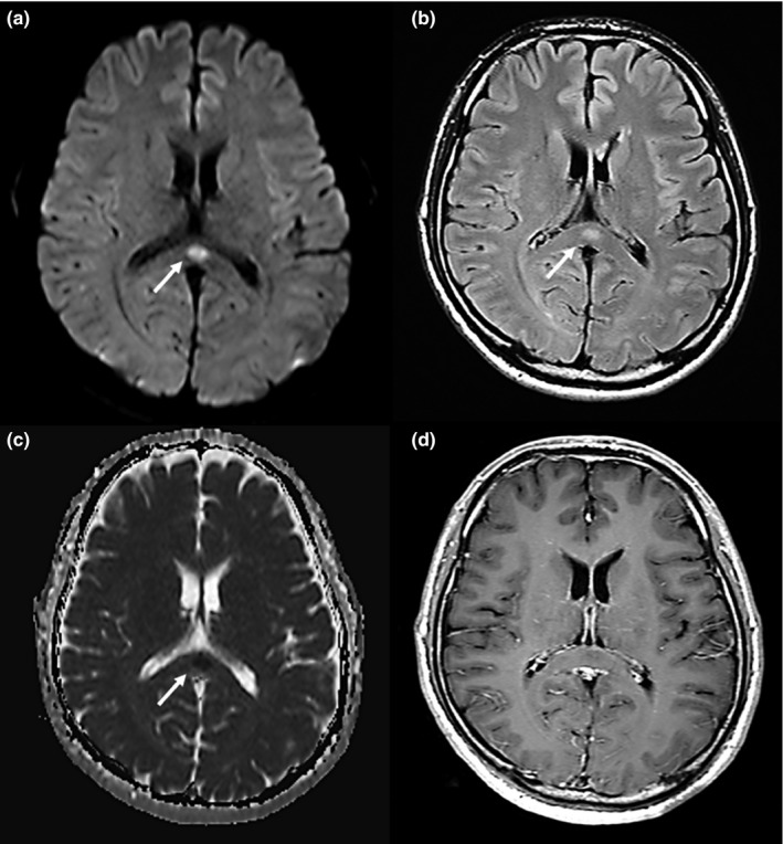
A 41‐year‐old man was admitted with fever, headache, and fatigue. He received a diagnosis of clinically mild encephalitis. Magnetic resonance imaging was performed on day 1 hospitalization. Diffusion‐weighted (a) and fluid‐attenuated inversion recovery (b) imaging showed a marked hyperintense signal (arrows) on the splenium of the corpus callosum (SCC). The apparent diffusion coefficient (c) showed a hypointense signal (arrow). T1‐weighted imaging with contrast material (d) showed an isointense signal that was not enhanced by the contrast material. Arrows indicate the SCC