
Figure 2.
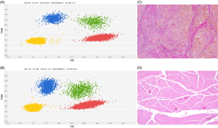
Genetic and pathologic findings in patients. A, B, Representative results from dPCR of DNA from ectopic muscle biopsies. Digital PCR quantifies the load of mosaic mutation, c.1624G > A (p.Glu542Lys), in DNA extracted from ectopic dorsal interosseous muscle and ectopic extensor digitorum muscle from patient 2. Blue cluster (FAM) shows signals from mutant allele, red signals (VIC) from reference allele and green signals from both mutant and reference alleles. Yellow cluster represents the wells where no amplification signal was detected. C, Histopathological findings from affected muscle in patient 1. Severe changes with increase in connective tissue (van Gieson). D, Histopathological findings from affected muscle patient 2. Scattered rounded eosinophilic muscle fibers in otherwise normal looking tissue (H‐E). Bars: 100 μm