Figure 5.

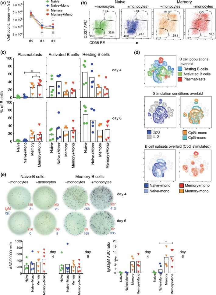
Monocytes do not enhance B‐cell subset differentiation when stimulated with CpG. (a) Count of cells in the cultures before and after stimulation with CpG. (b) FACS profiles of B cells from a representative donor that have been stimulated with sCD40L, IL‐21 and CpG, with and without monocytes for 4 days. (c) Percentages of B cells in the three analysis gates defined by CD27 and CD38 expression showing values for individual donors (symbols, n = 5) and means for all donors (bars). Asterisks indicate significance using paired t‐test, *P < 0.05, **P < 0.01. (d) tSNE plots indicate clustering of cells within the B‐cell gate of a representative donor assessed on day 6, concatenating data with and without CpG and/or monocytes, and overlaying B‐cell analysis gates based on CD27 and CD38 expression (top panels), stimulation conditions (middle panels) and the two subsets stimulated with and without monocytes (bottom panels). (e) Effect of monocytes on ASC detection after stimulation with CpG, as in Figure 3. ELISPOT images are shown for a representative donor, and ASC numbers, as well as ration of IgG to IgM ASCs, are shown for five donors. Asterisks indicate significance, as above. Results of five different donors from three independent experiments.
