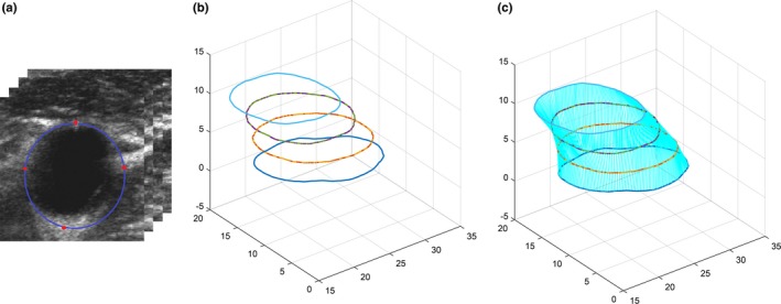
Figure 4.

The initialization of MAB segmentation. (a) User interaction on transverse slices of three‐dimensional ultrasound common carotid artery images every 3 mm (i.e., every three slices). The red points are located manually and the blue contours are generated using cubic spline interpolation. (b) Generated initial contours of all slices in (a). (c) Interpolated initial MAB contours from the corresponding points of the two adjacent user interaction slices in (b). [Color figure can be viewed at http://wileyonlinelibrary.com]
